Metabolic Imaging with RF Antennas to predict Chemotherapy Efficacy

An accessible and affordable virtual biopsy tool that can identify cancer biomarkers and tumor activity without shedding a drop of patient blood
Being able to predict the response of patients to chemotherapy would undoubtedly be beneficial for patient well-being, avoiding the unnecessary toxic side effects. The metabolites phosphoethanolamine (PE) and glycerophosphocholine (GPC) are involved in cell membrane metabolism, and the PE to GPC ratio goes up during malignant transformation of cells. The EU-funded MIRACLE project aims to validate this ratio as a biomarker of predicting cancer treatment response. Scientists will assess these phospholipid biomarkers in patients in a non-invasive manner using MRI under different field strengths. The results of the study have the potential to bring the approach a step closer to regulatory clearance for clinical use.
Metabolic Imaging with RF Antennas to predict Chemotherapy Efficacy: We will validate the phospholipid biomarker that can predict response of chemotherapy in patients with breast cancer using the most powerful MRI system in the world. In particular the ratio of phosphoethanolamine (PE) to glycerophosphocholine (GPC), metabolites from the build up and breakdown of cell membranes, have been shown to increase accuracy in prediction of chemotherapy response from 75% to 96% [1]. At the clinically available field strength of 7T, we have recently demonstrated that we can image these phospholipid biomarkers non-invasively. In order to assess accuracy and bring the detection of the biomarker to FDA approval, we will spin out a validation study to investigate the accuracy in detecting the biomarker by comparing the results from the same subjects obtained at clinical MRI as well as at 10.5T MRI. The innovation idea is linked to the Non-Invasive-Chemistry-Imaging (NICI) project of FET-OPEN-01-2016-2017-801075. More specifically to the deliverable D6.1: “List of groups and stakeholders for targeted dissemination”. Here the potential of the NICI project that uses multi-nuclear 7T MRI in patients with metastasis in liver was shown to the stakeholders in breast cancer imaging (joint meeting of International Society of Magnetic Resonance in Medicine (ISMRM) with EUropean Society Of Breast Imaging (Eusobi) in Las Vegas 2018). The importance of imaging chemistry was immediately recognised by all medical doctors present (radiology, surgery and oncology), and fast track to FDA clearance was discussed. By establishing a small consortium of the UMC Utrecht that leads the project, a newly formed spin out company that provides the 7T and 10.5T specific hardware, and University of Minnesota that has access to patients and houses the world’s strongest MRI, we will conduct the biomarker validation in 10 subjects scanned at both systems.
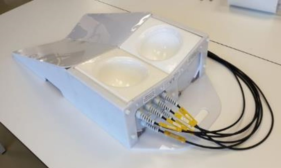
WaveTronica contributed to the MIRACLE project by engineering a mammacoil setup. To be able to do a comparison study on a 7tesla and 10.5tesla MRI scanner. Since these two MRI scanners inherently operate with different radio frequencies for both the proton and phosphorus nuclei imaging. The coils cannot be made identical. But for a fair comparison, the coils needed to be similar enough, I.e. meet minimum specifications. WaveTronica realized several test versions and mostly spent time on reorganizing parts (antenna like structures) on the same holder mechanics. During this process, the most used equipment is a vector network analyzer, but also, at some point, the MRI scanner from UMCU was needed to verify the findings from the bench measurements. A workable solution for 7tesla was found that could be translated to 10.5T. The final preproduction versions with a fitting holder were designed and shipped to the USA with accompanying documentation (including results from the safety study performed by the UMCU). These documentations are essential for the Minnesota University local safety commission for scanner clearance of this new hardware. To be able to compare scanner performance between UMCU and Minnesota scanners, a Phantom was designed by WaveTronica and shared with both parties.
Before WaveTronica can bring the MRI coil and the insight on cancer treatment progression this technology can bring to the patient. More work and studies need to be performed. As promising as this new technology is, and as much as our team would like to move forward. Medical technology development is time-consuming, expensive, and bound by regulations. To keep moving forward, WaveTronica has found interested investors and applied for a new EU grant in a new consortium. This project has been a significant investment for a small enterprise as WaveTronica. Nonetheless, we believe it is vital to the future of our company and also could be (partially) to the future of MRI worldwide as a tool to improve patient treatments.


